01
Viral vector tracing
AAV and rabies-virus tracing in Cre-driver mouse lines to map afferent and efferent connectivity of molecularly defined STN and dopamine subpopulations.
Methods
Our experimental approach combines genetic access to defined cell types, viral circuit tracing, optogenetic perturbation and high-resolution imaging — all anchored in carefully controlled mouse behaviour.
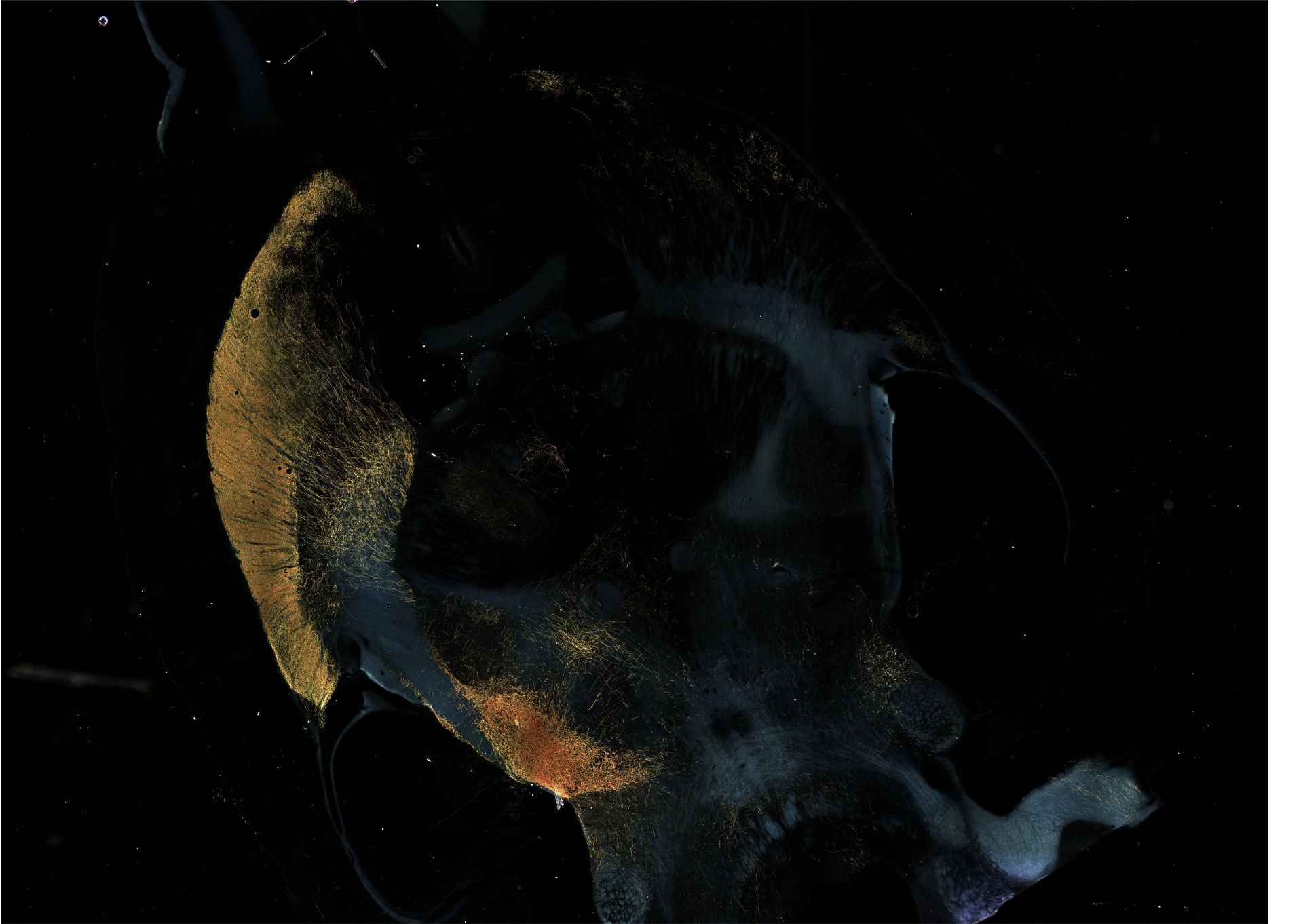
AAV and rabies-virus tracing in Cre-driver mouse lines to map afferent and efferent connectivity of molecularly defined STN and dopamine subpopulations.
Cell-type-specific activation and silencing using optogenetics and chemogenetic DREADDs (Gs, Gq, Gi, KORD), enabling precise, reversible control of genetically defined neurons.
Multiplexed FISH with post-FISH immunofluorescence (ORAMACELL protocols) to co-detect mRNA and protein in fresh-frozen sections, enabling identification of rare neuronal subtypes.
Quantitative motor, motivational and aversive learning assays in mice, including operant behaviour paradigms and the 6-OHDA model of Parkinson's disease with a refined survival protocol.
Image acquisition and quantitative cell counting of FISH-labelled cells across rostro-caudal extents of the STN, midbrain and forebrain.
Cross-species spatial transcriptomics and snRNA-seq to compare subthalamic and para-subthalamic populations between mouse, non-human primate and human tissue.
Amperometric biosensors to measure acute neurotransmitter and metabolite release in vivo: dopamine, glutamate, acetylcholine, GABA, lactate and pyruvate.
Custom algorithms for automated cell quantification of microglia and TH cells, plus 4D movement analysis for rats and mice.